Image 9
|
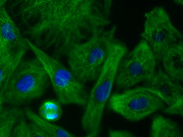
Image 9 |
| This image should be viewed in color. It represents the superposition of images 7 and 8. Each image was colored to correspond to the color seen in the microscope, and these two colored images were then combined. Blue represents the DAPI-stained chromatin and green represents the antibody-stained tubulin. One of the cells near the lower left corner of the picture is in mitosis. For further explanation, see the legend to Images 7 and 8. |
| Click on the image to return to the previous page. |